 |
 |
||||||||||
| MPE | ||||||
L - EDGE SPECTRA of Cs and Xe
Related publications: J. Padežnik Gomilšek, A. Kodre, I. Arčon, M. Hribar Phys. Rev. A 68 (2003) 042505
Abstract
X-ray absorption coefficient in the vicinity of L edges of Cs has been
measured on an alloy of Cs and Na. The absorption spectrum of the solid
sample is virtualy free of stractural signal, similarly to spectra
of monatomic gases. It exibits the same pattern of sharp multielectron
photoexcitation features as in the L edge absorption spectrum of the
adjacent element xenon.
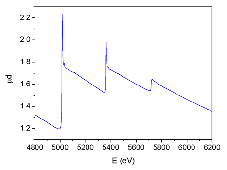 |
Fig.
1: The L-edge absorption spectrum of Cs measured on a thin
layer of the Na/Cs alloy. |
Introduction
The study of x-ray atomic absorption, measured either directly on a
monatomic gas sample [1-5] of an element or derived
from an x-ray absorption spectrum of a compound sample after removal
of the structural
signal [6-10] (EXAFS = extended x-ray
absorption fine structure), provides data on correlation in the
atomic system. The tiny sharp features on the
smoth energy dependence of the photoabsorption cross section are
fingerprints of multielectron photoexcitations (MPE). These reaction
channels arise from the change of the mean atomic field in the photoeffect
but also from the correlated motion of atomic electrons. A detailed
analysis can elucidate particularities in the coupling scheme and
configuration interaction of the atom [1,2,4,5].
The atomic absorption spectrum is a very good approximation to
the atomic absorption background (AAB) in the XAFS structural
analysis.
Since the structural signal and MPE occupy the same spectral region
above a major absorption edge, the small MPE features, if unrecognized,
interfere with the interpretation of the structural signal leading
to errors in the structure parameters determined in the XAFS analysis
[8,9].
In recent studies a parallel analysis of K-edge MPE in neighbor
elements (Ar - K; Kr - Rb) was successfully introduced [4,5].
The method exploits the fact that the cores of the two neighbor
elements (noble
gas and
alkali metal) are largely the same, apart from the unit difference
in the nuclear charge, and an additional loosely bound electron
in the outer shell of the alkali metal atom. From the comparison of
MPE excitations it was possible to deduce that common features
in
the spectra
follow from a specific interaction of core configurations, while
the differences in the MPE features of the two elements stem from
the additional
coupling of the valence s electron.
In this study we compare L-edge MPE in neighbor elements Xe and
Cs. Complete L-subshell MPE spectra have already been measured
on Xe
gas2). On the other hand, pure Cs atomic L-edge spectrum has
not been measured
yet. Only the most prominent resonant MPE features have been
extracted from the Cs L-edge EXAFS spectra measured on amorphous
materials
[11,12,13]. A measurement of the full atomic Cs L-edge
absorption spectrum, comparable
in detail to that of Xe, promises another fruitful comparison MPE
study.
The L-edge absorption spectrometry on monoatomic Cs metal vapor
requires a very demanding and costly heat-pipe cell4). Instead
we measured
the absorption on a thin layer of Cs/Na alloy in the vicinity
of its melting
point (70° C), where the weak EXAFS signal can be removed numerically.
Surprisingly, the experiment showed that the strong disorder
in the alloy produces practically pure Cs atomic absorption
spectrum almost
without an EXAFS component.
Experiment
Cs/Na alloy with a concentration ratio of 1:9 was prepared. A small
amount of the alloy was placed, together with a drop of paraffine
oil to prevent oxidation, into a small lucite container between
two kapton
foils and squeezed into a thin layer. The container and the oil kept
the metal perfectly stable for several hours of the experiment: no
sign of oxidation was observed after demounting. The stability of
the sample was confirmed also by the perfect reproducibility of
the scans
that were recorded in sequence at each of the L subshell edges.
The absorption experiment was performed at the E4 station of the
DORIS ring at HASYLAB synchrotron facility, DESY (Hamburg, Germany).
The
beamline provides a focused beam from Au-coated mirror and a Si(111)
double-crystal monochromator with 0.8 eV resolution at Cs L3-edge.
Harmonics are effectively eliminated by a plane Au coated mirror
and by detuning the monochromator crystal using a stabilization
feedback control. Exact energy calibration was established
with a simultaneous
measurement on a Ti metal foil (EK = 4966 eV) between the second
and
the third ionisation cell.
 |
 |
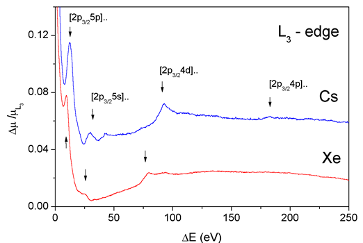 |
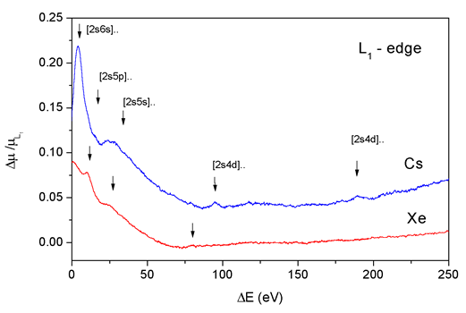
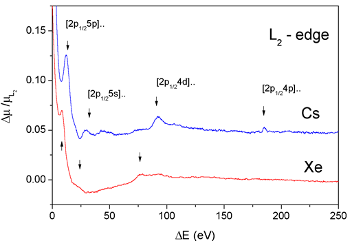
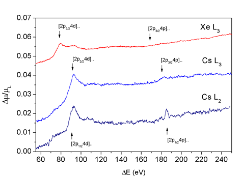
Fig.
2: The comparison of the absorption in the L subshell regions
of Cs and Xe after removal of the average trend to enhance
the detail. Theoretical estimates of the energy of double excited
states are shown by arrows. For each subshell, the origin of
the energy scale is shifted to the respective ionization threshold. |
Results
A compound picture of the L edges (Fig. 1) is obtained as a superposition
of three scans per subshell region. Each of the edges is preceded by
a resonance due to the excitation of the 2p or 2s electron to the unoccupied
bound states just below the continuum.
The spectra above each of the edges are remarkably flat, almost
without oscillatory EXAFS signal characteristic of solid samples.
A slight
convexity of the spectrum above the L3 and L2 edge, observed already
in the L absorption spectra of some heavy elements, has been explained
as a consequence of subshell polarization14).
The strongest of the sharp MPE features are just barely visible
in the spectrum. To expose the details, the average trend of each
subshell
region is removed from the relative cross section (Fig. 2). Several
groups of MPE can be clearly discerned above each absorption edge.
The groups can be identified by their energy as multiple excitations
involving electrons in consecutively deeper subshells from 6s to
4p, in complete analogy with the MPE groups in the neighboring Xe2),
shown
below. Hartree-Fock estimates [15] of the threshold
energies of the corresponding double excitations are indicated
by arrows.
Even at this level of magnification the oscillatory structural
signal is visible only close to the edge. The amplitude of EXAFS
oscillations
is smaller than the MPE features and vanishes in the noise level
about 80 eV above each edge.
The marked difference between MPE in Cs L1 and L2,3 spectra point
to the same orbital-momentum-sensitive mechanism of multielectron
coexcitation
as already established in Xe2). Closer inspection of the Cs L2
and L3 absorption spectra shows similarities in [2p4d] and [2p4p]
MPE features,
and reveals differences in their amplitude and shape compared
to those in Xe L3,2 spectra (Fig. 3). Differences may be ascribed
to resonant
and shake-up transitions into specific final states of a bound Cs
atom.
 |
| Fig. 3: The comparison of the [2p4d] and [2p4p] MPE features above Cs L2 and L3 and Xe L3 edges spectra. |
Acknowledgments
Support by the Ministry of Science and Technology of the Republic of
Slovenia, and by Internationales Buero BMBF (Germany) is acknowledged.
K. V. Klementiev of HASYLAB provided expert advice on beamline operation.
1. S. J. Schaphorst et al, Phys. Rev A 47, 1953 (1993).
2. I. Arčon et al, Phys. Rev. A 51 147 (1995).
3. R. Prešeren et al, Nucl. Inst. Meth. Phys. Res. B 111 161 (1996).
4. J. Padežnik Gomilsek et al, Phys. Rev. A 64 22508 (2001).
5. A. Kodre et al. J. Phys. B (submitted)
6. G. Li, F. Bridges, and G. S. Brown, Phys. Rev. Lett. 68 (1992) 1609.
7. A. Filipponi and A. Di Cicco, Phys. Rev. A 52 (1995) 1072.
8. P. DAngelo et al, Phys. Rev. A 47 2055 (1993).
9. A. Kodre, I. Arčon, R. Frahm, J. Phys. IV France 7, C2 195 (1997)
10. R. Prešeren et al, J. Synch. Rad. 8 (2001) 279.
11. A. Kodre et al, J. de Physique C-9, 4, 397 (1994).
12. A. Kodre et al, Physica B 208&209, 379 (1995).
13. J. A. Solera, J. Garcia, M.G. Proietti, Phys. Rev. B 51 2678 (1995-I).
14. W. Jitschin et al, Phys. Rev. A 35 5038 (1987).
15. C. Froese-Fischer, Comput. Phys. Commun. 43 355 (1987).
|
|
|||||||||||||||||||||||||||||||||||||||||||||||||||||||||||
|
E-mail:iztok.arcon@p-ng.si Last change: 28-Jun-2006 |
|||||||||||||||||||||||||||||||||||||||||||||||||||||||||||